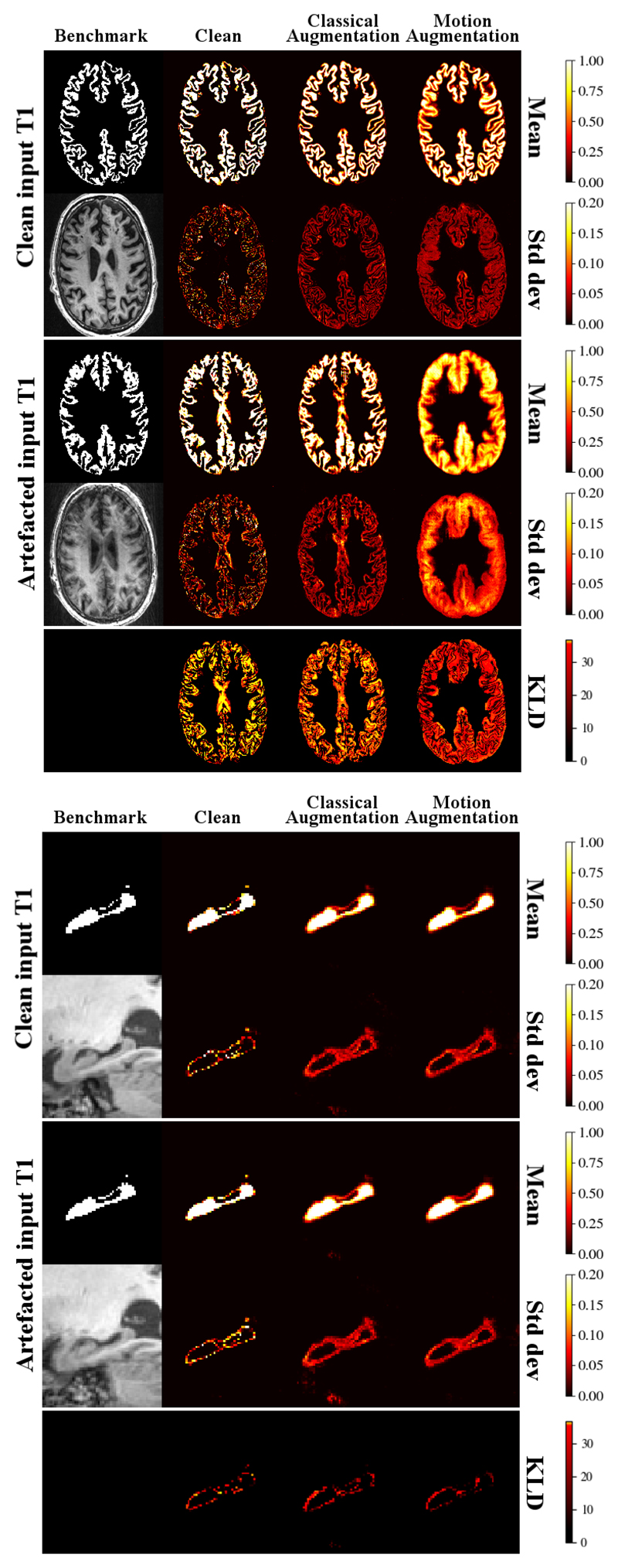
Fig. 6.
Per-voxel mean and uncertainty estimations on CGM (top) and hippocampus (bottom) segmentation tasks for clean (no augmentation), classically augmented and motion augmented models for a test-retest pair for which one scan is heavily artefacted. The segmentation produced by a benchmark method is shown for reference. Bottom row of each block: KL-divergence (KLD) between the probability distributions produced by each model on clean and artefacted scans.

