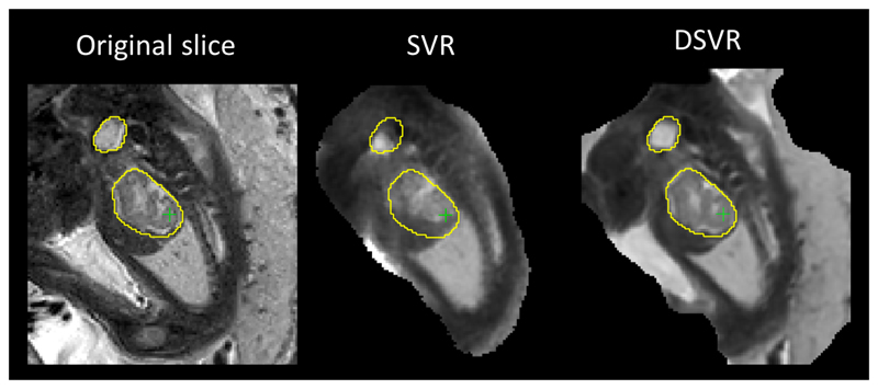
Fig. 7.
Comparison of SVR and DSVR in presence of non-rigid motion: original acquired slice (Yk) vs. slices simulated from SVR and DSVR (Ȳk). The yellow isolines delineate the structure in the original slice, and show misalignment with SVR reconstruction due to limitation of rigid motion correction. The problem is resolved by non-rigid motion correction in DSVR.

