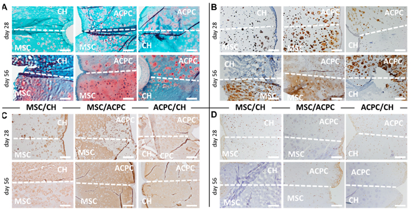
Fig. 7.
Histological sections of the layered co-cultures showing (A) safranin O staining for sGAGs, (B) collagen type II, (C) collagen type I, and (D) PRG4. The dotted line marks the interface between the two different cell-laden layers of the hydrogels, and the cell type residing in each zone is specified in overlay. Scale bar is 500 μm.

