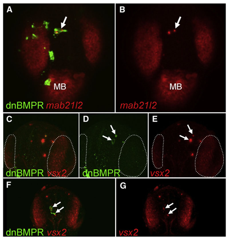
Figure 6. Ectopic Expression of Retinal Markers in BMP-Depleted Telencephalic Cells at 24 hpf.
(A-G) Embryos were injected with HSP70:dnBMPr plasmid at one- to two-cell stage followed by heat shock at oblong/early blastula stage. The arrows indicate dnBMPr-positive cells in the telencephalon at 24hr, which ectopically express retinal markers mab21l2 (A and B) and vsx2 (C-G). The number of transiently transgenic dnBMPr cells is significantly lower at 24 hpf than at earlier stages, and the remaining transgenic cells frequently undergo apoptosis (arrows in D). (C-E) White dotted lines mark the vsx2-positive retina. All views are dorsal, anterior at the top. All figures are z-projections of confocal sections. MB, midbrain. See also Figure S6.

