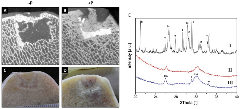
Fig. 4.
Micro-CT and macroscopic pictures, respectively, of implantation sites at 6 months for −P (A, C) and +P (B, D) scaffolds. XRD analysis of 3D printed CaP scaffolds (E); I indicates a sample before implantation, whilst II and III indicate samples at necropsy. The most prominent peaks are labelled as: B: brushite (PDF-No. 09-0077), M: monetite (PDFNo. 09-0080), T: β-tricalcium phosphate (PDF-No. 09-0169) and HA: hydroxyapatite (PDF-No. 09-0432).

