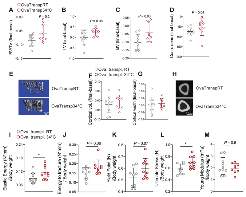
Figure 4. Warm–Microbiota Transplantation Prevents Bone Loss and Improves Bone Strength.
(A-E) Delta between two Micro-CT measurements (day 0 and day 32 after starting the microbiota transplantation) of proximal tibias at trabecular level in 21 weeks old ovariectomized, microbiota recipient female mice. The recipient mice were ovariectomized at week 16, and repetitivelly transplanted with fecal microbiota from 34°C exposed, or RT-kept donors (OvaTransp34°C, or OvaTranspRT, respectively). The 34°C treatment of the 16 weeks old female donor mice was initiated one month before the starting the transplantations, and lasted for the whole length of the experiment. Bone volume/tissue volume (BV/TV) (A), bone volume (BV), (B) total volume (TV) (C), and connectivity density (D). (E) Representative reconstruction (each consisting of 262 sections, n = 10 per group) of trabecular bone use for the calculations. Scale: 100μm.
(F-H) Cortical bone volume (F) and width (G) measured in midshaft of tibias from mice as in (A). (H) representative cortical section (from 62 sections per bone of each mouse of n = 10 per group), scale: 0.5mm.
(I-M) Biomechanical analysis of tibias from mice as in (A) showing elastic energy (I), energy to fracture (J), yield point (K), ultimate stress (L) and Young’s modulus (M), all normalized to their body weights. Data are shown as mean ±SD (n = 10 per group). Significance is calculated using Mann-Whitney t-test *P < 0.05; **P < 0.01; ***P < 0.001.

