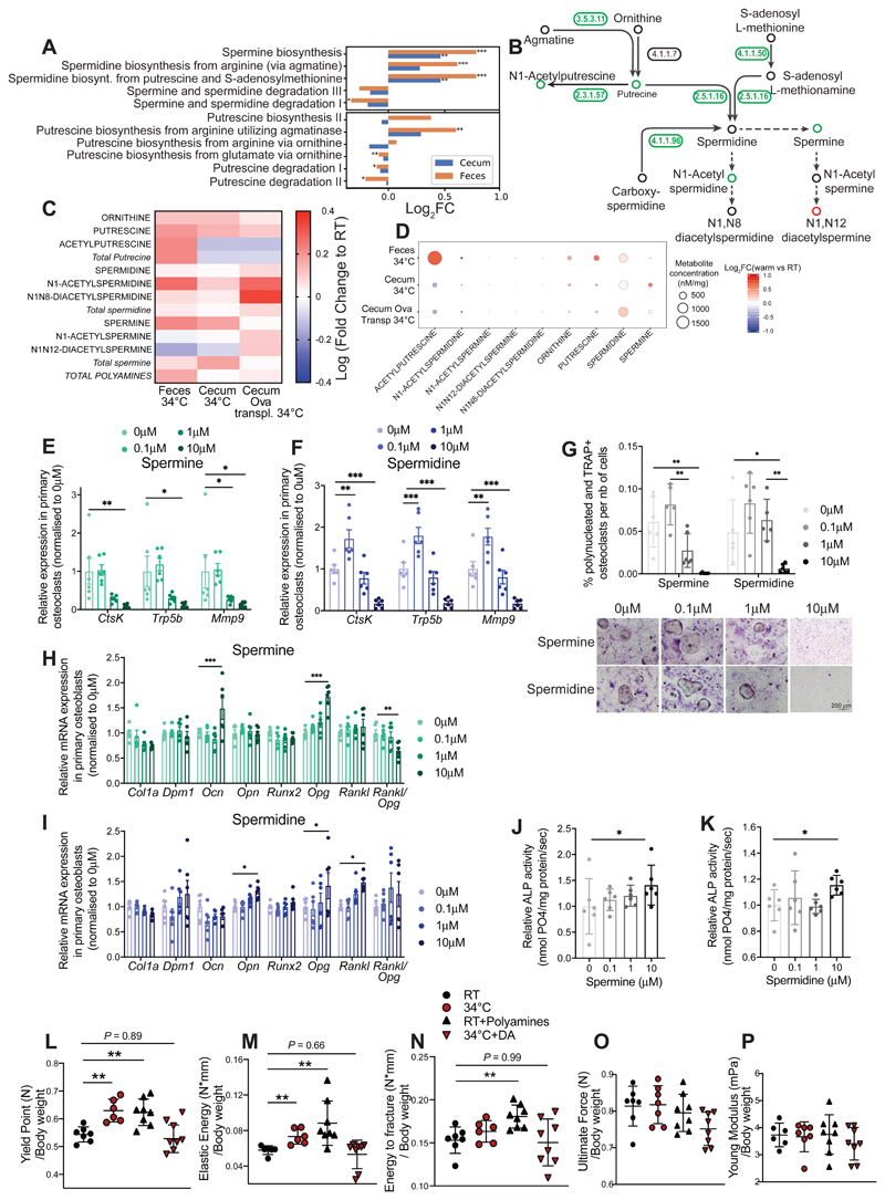
Figure 7. Microbial Production of Polyamines Mediate the Warmth Effects on the Bone.
(A) Bar chart representing metagenomics analysis of the bacterial polyamine biosynthetic and degradation pathways in feces and cecum samples from 24 weeks old female mice that were exposed to 34°C for 2 months, or kept at RT. Significance shows false discovery rate (FDR): *P < 0.05; **P < 0.01; ***P < 0.001.
(B) Graphical representation of the polyamine biosynthetic pathway where numbers (enzymes from keg nomenclature) represent the level of respective genes present in the gut microbiota from fecal samples of mice as in (A). Green–coloured circles show increase, red–coloured show decrease, and black–coloured indicate unchanged concentrations after warm exposure. 3.5.3.11: agmatinase, 4.1.1.7: benzoylformate decarboxylase, 2.3.1.57: putrescine acetyltransferase / spermine-spermidine N1-acetyltransferase, 4.1.1.50: adenosylmethionine decarboxylase, 2.5.1.16: spermidine synthase, 4.1.1.96: carboxynorspermidine decarboxylase.
(C and D) Heatmap (C), or heatmap associated with absolute polyamine levels (D), showing fold change of polyamines measured using hydrophilic interaction liquid chromatography coupled to tandem mass spectrometry (HILIC - MS/MS) in feces or cecum samples from 24 weeks old female mice that were exposed to 34°C for 2 months versus RT controls (34°C vs. RT; Feces 34°C and Cecum 34°C); or cecum of 21 weeks old ovariectomized, microbiota recipient female mice (Cecum Ova transpl34°C).
(E and F) Relative mRNA expression levels in cultured primary osteoclasts subjected to different spermine (E) or spermidine (F) concentrations, measured by qPCR.
(G) Quantification of the polynucleated and TRAP+ differentiated osteoclasts normalized to the total number of cells in presence of spermine or spermidine. (Below) Representative images (from 6 wells per condition) from TRAP staining of osteoclasts differentiated in presence of spermine or spermidine. Scale: 200 μm.
(H and I) Relative mRNA expression levels in cultured primary osteoblasts subjected to different spermine (H) or spermidine (I) concentrations, measured by qPCR.
(J and K) Relative alkaline phosphatase (ALP) activity in osteoblast culture after spermine (J) or spermidine (K) supplementation at different concentrations.
Significance (p-value) in all panels except (A and L-P) is calculated using Mann-Whitney t-test: *P < 0.05; **P < 0.01; ***P < 0.001.
(L-P) Biomechanical analysis using 3–point bending test of femur from 23 weeks old female mice that were either RT kept (RT); warm exposed (34°C); supplemented with freshly prepared polyamine mix and RT kept (RT-Polyamines); or provided with 50μm Diaminazene Acetureate (DA) and kept at 34°C (34°C-DA), starting at 16 weeks of age until sacrifice. Polyamines and DA were supplemented in drinking water every second day. The panels show yield point (L), elastic energy (M), energy to fracture (N), ultimate force (O), and Young’s modulus (O) that are normalized to their bodyweight values at sacrifice.
Data are shown as mean ±SD (n = 8 per group). Significance is calculated based on One-Way ANOVA: *P < 0.05; **P < 0.01; ***P < 0.001.

