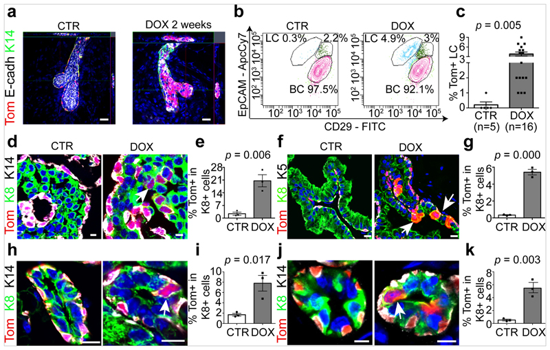
Fig. 1. LC ablation promotes BC multipotency in glandular epithelia.
a, Whole-mount confocal imaging of immunostaining for tdTomato (Tom), K14 and E-cadherin (E-cadh) in the mammary glands of K5CreER/tdTomato/K8rtTA/TetO-DTA mice after IDI with NaCl (Control, n = 3 independent experiments) or 0.2 mg DOX (n = 3 independent experiments) and chasing for two weeks. Scale bars, 20 μm. b, Representative FACS plot of CD29 and EpCAM expression in Lin−tdTomato+ epithelial cells from control mice (n = 5) and from mice 1 week after DOX administration (n = 16). The percentage of the gated population out of all epithelial cells is shown. c, Quantification of tdTomato expression in CD29low EpCAMhigh LCs from control (n = 5) and DOX-treated (n = 16) mice. The bar height and error bars are mean ±s.e.m., with individual data points shown. P values are derived from unpaired two-sided t-tests. d–k, Confocal imaging of immunostaining for tdTomato, K8 and K14 or tdTomato, K8 and K5 in the MG organoid (d), prostate (f), salivary gland (h) and sweat gland (j) tissues and the respective quantification of tdTomato+ cells in K8+ LCs (e, g, i, k) in K5CreER/tdTomato/K8rtTA/TetO-DTA organoids (d, e) or in mice (f–k) treated with DOX and analysed 72 h later for organoids and 1 week later for in vivo mouse experiments. Scale bars, 10 μm (organoid); 5 μm (mouse tissues). Hoechst nuclear staining is shown in blue in immunofluorescence images. n = 3 independent experiments. In e, g, i, k, the bar height and error bars are mean ±s.e.m., with individual data points shown. P values are derived from unpaired two-sided t-test.

