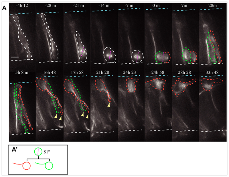
Fig. 4. Symmetric neuron production.
(A) A terminal division producing two neurons. The nucleus of a cell (white dashed outline) migrates apically (white broken line) and divides. The cleavage plane (pink broken line) is perpendicular to the apical surface. Following mitosis the two daughter cells (red and green dashed outlines) maintain apical attachment and their nuclei migrate back towards the basal surface (blue broken line). Once the nuclei are at the basal surface both apical processes withdraw. The nucleus of the cell outlined in green becomes obscured, but three-dimensional analysis confirms that it also continues to withdraw its apical process (yellow arrowhead). Scale bar: 10 μm. Each image is a MIP through 30 z-sections imaged at 1.5 μm intervals. (A′) Lineage tree for A. (See Movie 4 in the supplementary material.)

