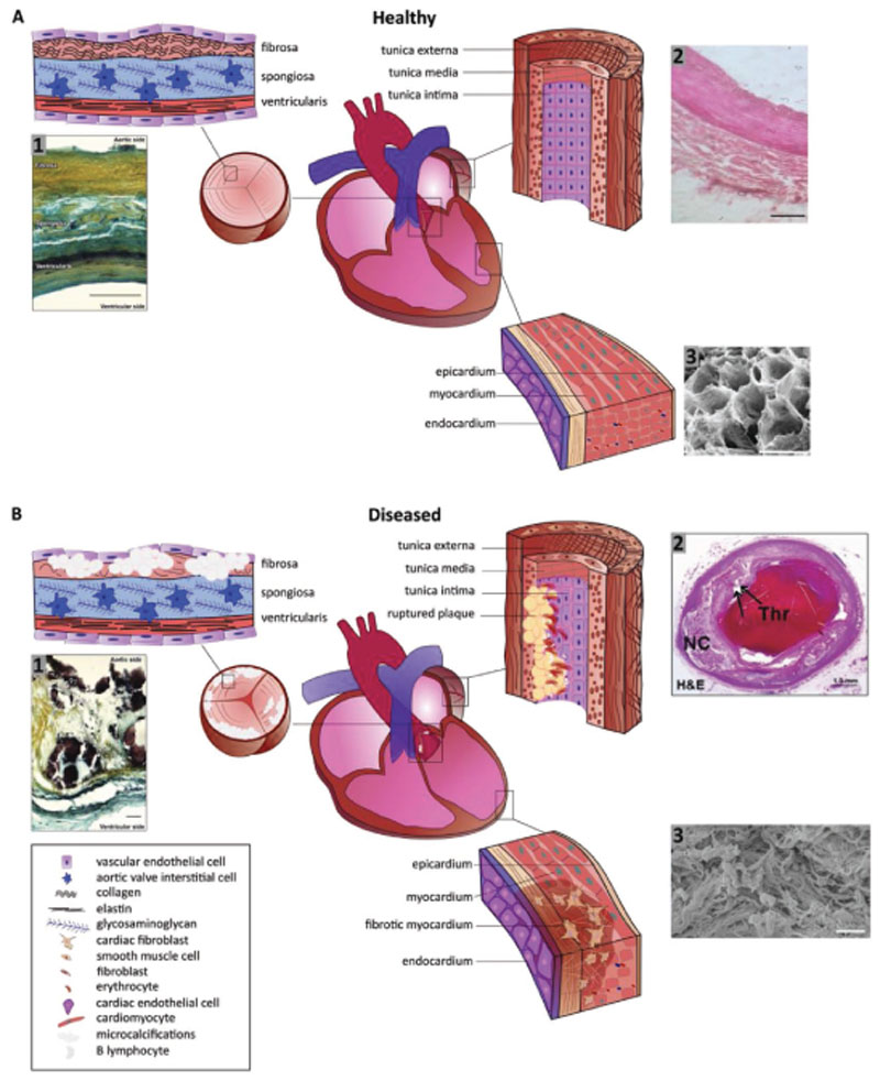
Figure 1.
Schematic representations of A) the healthy heart with enlargements of the aortic valve highlighting the layer composition, of the coronary arteries outlining the fiber orientation in the tunica, and of the myocardial wall with cellular and fiber direction, and B) the diseased heart illuminating fiber disruptions in the aortic valve due to (micro)calcification, in the coronary arteries due to plaque formation, and in the myocardium due to fibrosis. A1) Movat’s pentachrome staining of structural organization in a circumferential cross-section of a human aortic valve leaflet. Yellow = collagen, blue = glycosaminoglycans (GAGs), black = elastin, dark brown/black = calcification. Trilayered structure of the fibrosa (collagen-rich), spongiosa (GAG-rich), and ventricularis (elastin-rich) layers (scale bar = 100 μm). Reproduced with permission under the terms of the CC BY 4.0 license.[194] Copyright 2018, the Authors, Published by MDPI. A2) Histology section of coronary arterial wall highlighting the fiber structure and orientation (scale bar = 10 μm). Reproduced with permission under the terms of the CC BY 4.0 license.[195] Copyright 2018, the Authors, Published by MedWin Publishers. A3) Scanning electron microscopy photograph displaying the honeycomb-like structure of supportive fibers in the myocardium cross-section (scale bar = 50 μm). Reproduced with permission.[43] Copyright 2016, JoVE. B1) Movat’s pentachrome staining of structural organization in a circumferential cross-section of a human aortic valve highlighting the disruption of the leaflet layers by calcifications (black) and fibrosis (yellow regions) in CAVD (scale bar = 50 μm). Reproduced with permission under the terms of the CC BY 4.0 license.[194] Copyright 2018, the Authors, Published by MDPI. B2) Histology section of coronary arterial wall highlighting the plaque rupture with acute luminal thrombus (Thr) and underlying large necrotic core (NC). Arrows indicate the site of fibrous cap disruption (scale bar = 50 μm). Reproduced with permission.[196] Copyright 2013, Elsevier. B3) Scanning electron microscopy photograph showing the randomly arranged bundles of collagen fibres after myocardial infarction and myocardial scarring inside left ventricle (scale bar = 10 μm). Reproduced with permission.[197] Copyright 2017, Springer.

