Figure 3.
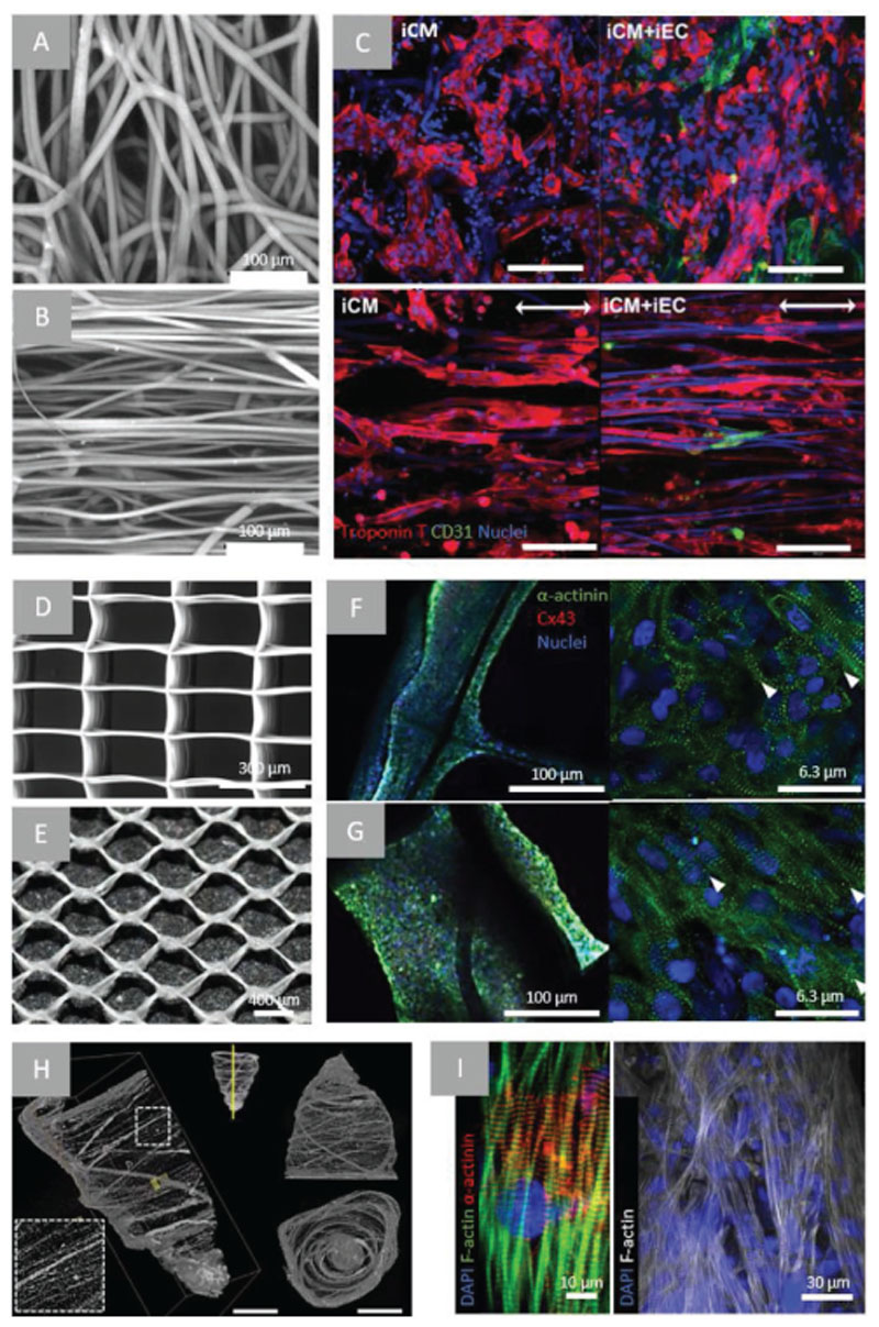
Examples of microfiber scaffolds applied for myocardial TE approaches. A) confocal microscopy images of PCL electrospun mesh with randomly oriented fibers and B) aligned fibers following heat and mechanical manipulation, C) confocal microscopy of iPSC-CMs (troponin T—red) and/ or iPSC-ECs (CD31—green) after 48 h of culture on random or aligned PCL electrospun scaffolds (scale bar = 100 μm). Arrow indicating direction of fiber alignment. Reproduced with permission. Copyright[167] 2017, The Royal Society of Chemistry). D) SEM images of PCL scaffold fabricated using MEW of rectangular (150 × 300 μm)[117] and E) hexagonal pattern (hexagon side length = 400 μm), confocal microscopy images of a-actinin (green), nuclei (blue) and connexin-43 (red) staining of iPSC-CMs in rectangular (F) and hexagonal (G) scaffolds.[34] H) μCT of pull-spun rat ventricle scaffold (scale bars: left = 10 mm, right = 5 mm), I) immunofluorescent stainings (as indicated) of iPSC-CMs cultured in scaffolds for 14 days. Reproduced with permission.[173] Copyright 2018, Springer Nature.

