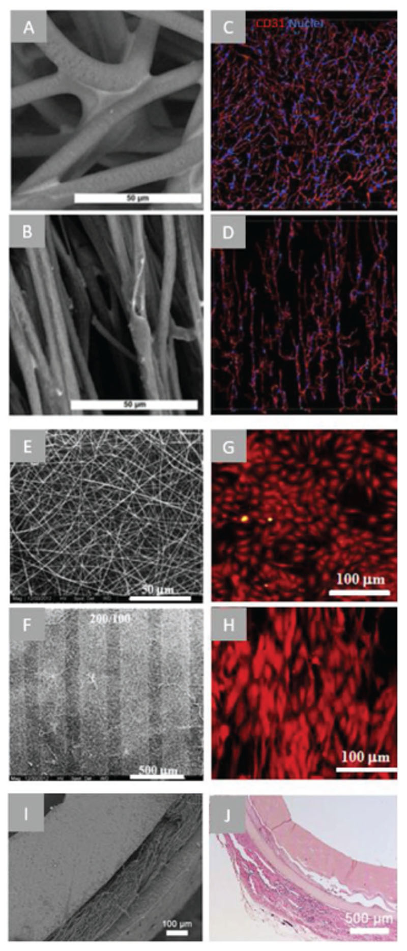
Figure 5.
Examples of fibrous scaffolds applied on the vascular engineering. A) SEM images of a random, B) and aligned electrospun scaffold (scale bar = 50 μm), C) CD31 staining (red) and DAPI staining (blue) generated configurations of iPSC derived endothelial cells on a randomly oriented scaffold and D) an aligned electrospun scaffold. Scale bars not stated in original paper. Reproduced with permission. Copyright[182] 2017, Springer Science+Business Media. E) SEM pictures of a random electrospun fibrous scaffold and F) a patterned electrospun scaffold with the ridge/groove width of 300/100 μm, G) phalloidin staining of endothelial cells loaded on the random electrospun scaffold and H) smooth muscle cells on the patterned scaffold showing a preferred direction of smooth muscle cell actin fiber morphology on the patterned scaffold. Reproduced with permission.[183] Copyright 2015, Elsevier. I) SEM image of the cross section a trilayer tubular graft obtained by three-step electrospinning, J) H&E staining of cross section of transplanted constructs 2 weeks post transplantation in mice. Reproduced with permission.[185] Copyright 2018, Elsevier.

