Fig. 3.
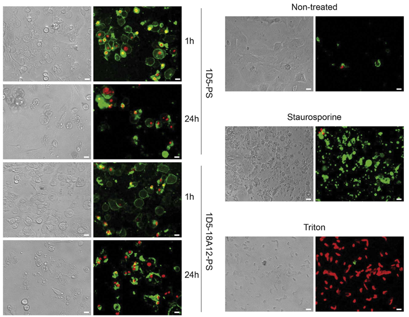
Nanobody-targeted PDT induces changes in morphology and cell death. HER2-positive HCC1954 were incubated with 25 nM nanobody-PS, 1D5-PS (upper left row) and 1D5-18A12 (lower left row), followed by light illumination (10 J/cm2 of light dose). Apoptotic cells were detected with Annexin V-FITC (green), whereas necrotic cells with propidium iodide (red) staining 1 h and 24 h after light illumination. The increase of propidium iodide staining shows the toxic effect of PDT increased between 1 h and 24 h. As a control for apoptotic and necrotic cell death, non-treated HCC1954 cells were incubated with Staurosporine or Triton respectively (right row). Pictures were obtained with an EVOS Microscope equipped with 10× objective. Scale = 20 μm. (For interpretation of the references to colour in this figure legend, the reader is referred to the web version of this article.)

