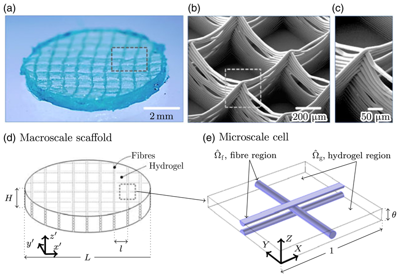
Figure 1.
(a) Optical microscope image of a fibre-reinforced hydrogel with a square fibre lattice of 800 μm. Note that the overall dimensions of the construct shown here are slightly different to those used in later experimental comparison. (b) Scanning electron microscopy (SEM) image of the fibre scaffold prior to it being cast in the hydrogel. (c) SEM image showing a detail of fibre buildup at the interconnection between printed vertical layers. (d) Schematic diagram of the idealised scaffold used in the homogenised model of this paper. (e) Schematic diagram of the microscale repeating cell, showing the microscale hydrogel region Ω̂g, and the microscale fibre region Ω̂f. The characteristic length scale at the microscale is the horizontal fibre spacing l, and the characteristic macroscale length is the overall diameter of the scaffold L. It is assumed that the scaffold diameter is much greater than the fibre spacing and that their ratio ε = l/L ≪ 1, which permits a separation of length scales as described in Section 2.3.

