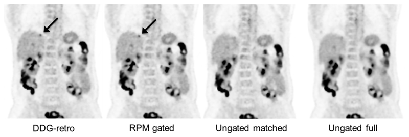
Figure 5.
A coronal slice showing an 18F-FDG avid liver metastasis (indicated by arrow) which is easier to detect and has a higher SUVmax on the two gated reconstructions, as compared to ungated. In this example, the DDG-retro and RPM-gated images received an equal score for overall image quality, and both were considered superior to the ungated images. The lesion indicated by the arrow was not considered to be definitely visible on the Ungated matched image, and was borderline-visible on the Ungated full image. One can also see the reduction in noise in the Ungated full image, as compared to the other three images. The images are on an SUV grayscale of 0–6.

