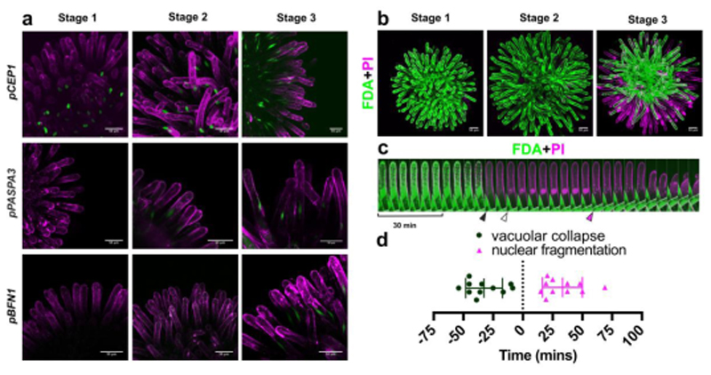
Figure 2. Ageing unpollinated papilla cells die in a dPCD-like process.
a) Maximal projections of confocal microscopy images showing promoter-reporters of canonical dPCD-associated genes, which are expressed during stigma senescence. Green represents the nuclear-localized H2A-GFP reporter; magenta shows propidium iodide (PI) staining the cell wall. b) FDA (green) and PI (magenta) staining in different stages of stigma senescence shows loss of cellular viability in peripheral papilla cells at stage 3 by absence of FDA staining, and entry of PI into the dying cells. c) Kymograph of an individual papilla cell stained with FDA and PI, showing an attenuation of the FDA signal and vacuolar collapse (black arrowhead), followed by entry of PI in the nucleus (white arrowhead), abrupt nuclear fragmentation (magenta arrowhead), and cellular collapse. d) Quantification of the timing of vacuolar collapse and nuclear fragmentation in relation to PI entry into the nucleus (time point ‘0’). Scatter plot with mean±SD, n=11 cells from three different stigmata. Mean time of vacuolar collapse is -32.59 ± 15.44 minutes, mean time of nuclear fragmentation is 33.32 ± 16.8 minutes. Scale bars, 50 µm.

