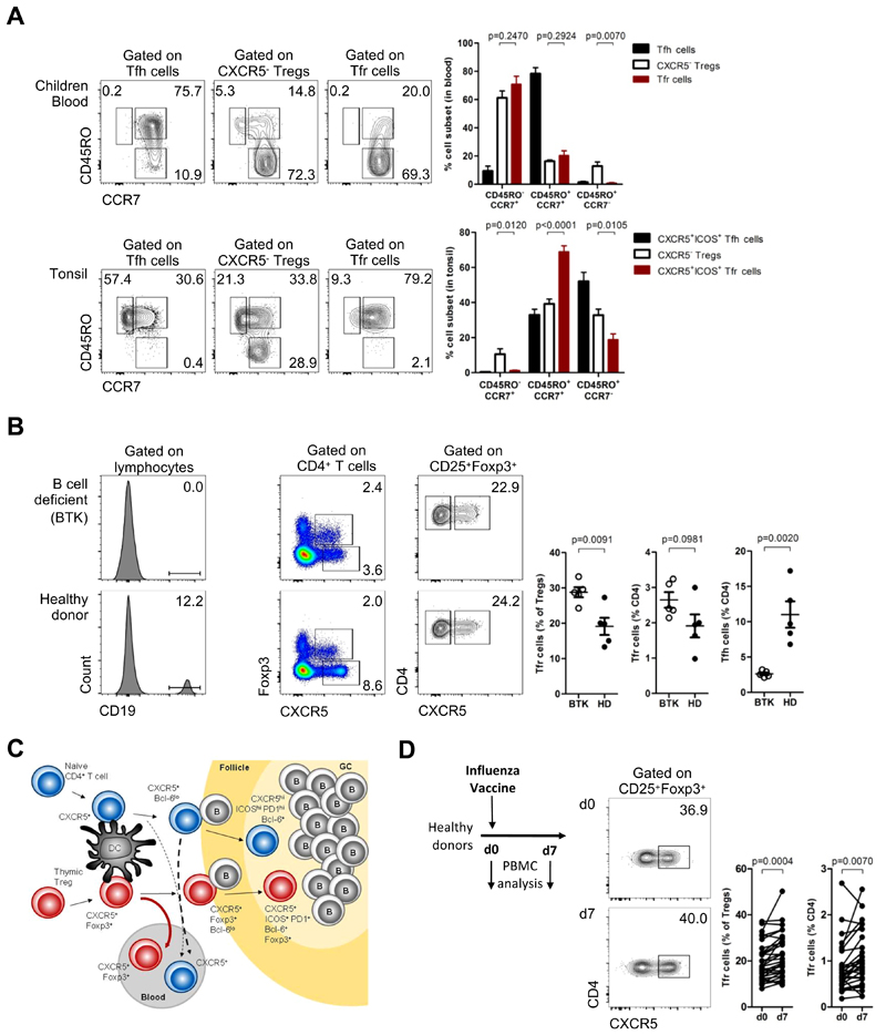
Figure 6. Blood Tfr cells are lymphoid tissue derived Tfr precursors.
(A) CD45RO+CCR7- EM, CD45RO+CCR7+ CM and CD45RO-CCR7+ naïve subsets of Tfr cells and CXCR5- Tregs in children blood (top) and in tissues (bottom). Representative plots (left) and pooled data (right) (n = 6, Student t-test). Tfh cells are represented in blue, CXCR5- Tregs in black and Tfr cells in red. CXCR5+ subsets in tonsils were defined as CXCR5+ICOS+ cells (Fig. S2B). (C) Blood Tfh and Tfr cells from X-linked Agammaglobulinemia (BTK-deficient) patients, compared to sex and age-matched healthy donors. Representative plots (left) and pooled data (right) (n = 5, Student t-test). (C) Model of CXCR5+ follicular helper and regulatory cells T cells generation and recirculation in humans, upon antigen stimulation. Tfh cells in red and Tfr cells in blue. (D) Frequency of peripheral blood Tfr cells on the day of influenza vaccination (d0) and 7 days later in healthy volunteers. Schematic representation and representative plots (left) and pooled data (right) (n = 32, Student t-test). Bars represent SEM. significant).

