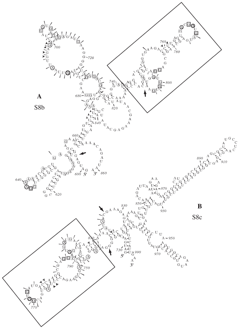
Fig. 6. Summary of RNA structure probing results for S8b and S8c.
Structure probing was carried out on panel (A) S8b (nt 596–861) and (B) S8c (728–993). The sensitivity of bases to the various probes is indicated by a black arrow head (double-stranded RNA—RNase CV1), circles (single-stranded A—RNase U2), boxes (single-stranded G—RNase T1) or a slender arrow (single-stranded RNA—lead acetate). The imidazole probing data, similar to that seen with lead acetate, are omitted for clarity. Large arrows represent regions of the predicted structure where structural information can be assigned. A conserved structure motif shared between these NS2-binding RNAs was identified (boxed).

