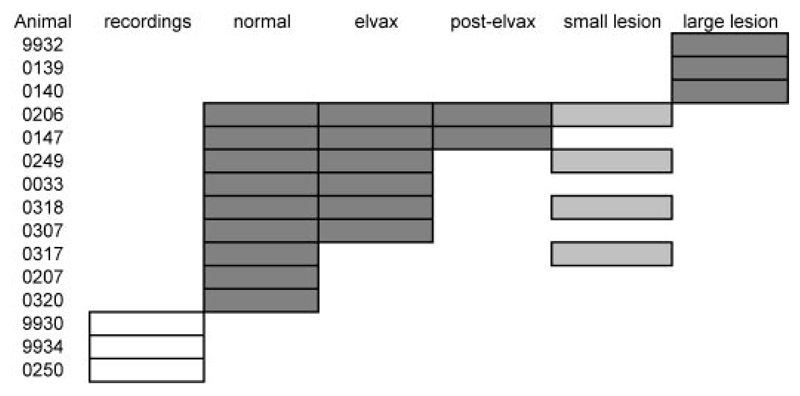
Fig. 1.
Summary of animals used in this study. Gray boxes indicate that an animal was tested for its ability to localize sound in the midsagittal plane in the condition indicated. For example, animal F0206 was trained and tested at all stimulus durations, providing control data (“normal”). These measurements were then repeated with 40-ms noise bursts after inactivating primary auditory cortex (A1) bilaterally with muscimol-Elvax (“elvax”), again after Elvax removal (“post-elvax”), and once more after making bilateral aspiration lesions of A1 (“small lesion”); “large lesion” refers to animals in which more extensive regions of auditory cortex were removed bilaterally. Dark gray boxes indicate that the animal was used for behavioral testing only, whereas light gray boxes (i.e., all A1 small lesions) indicate that electrophysiological recordings were made as well, before aspirating the cortex. Empty boxes indicate that the animal was used for electrophysiological recordings only.

