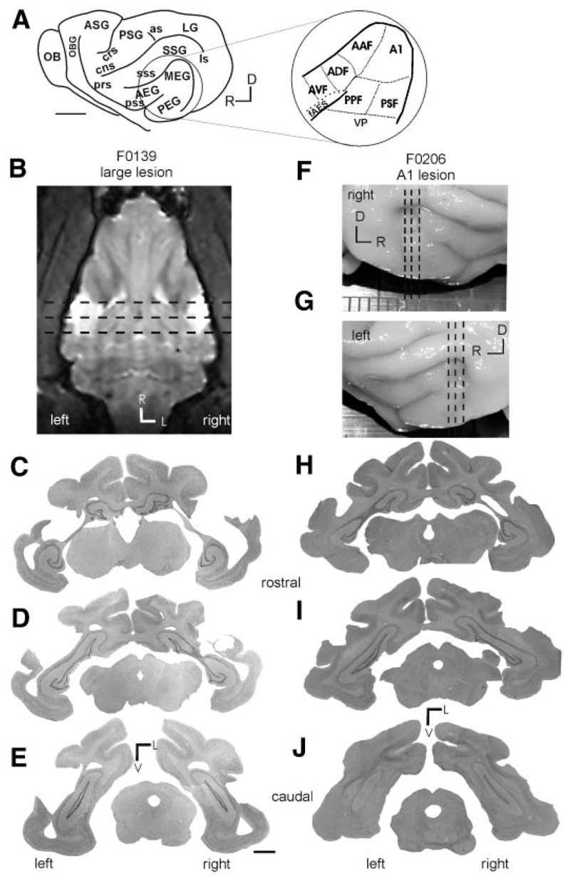
Fig. 4.
Location of the cortical lesions in 2 of the ferrets (0139 and 0206) used in this study. A: schematic of a ferret brain illustrating the location of the major gyri (OB, olfactory bulb; OBG, orbital gyrus; ASG, anterior sigmoid gyrus; PSG, posterior sigmoid gyrus; LG, lateral gyrus; SSG, suprasylvian gyrus; MEG, middle ectosyslvian gyrus; PEG, posterior ectosyslvian gyrus; AEG, anterior ectosyslvian gyrus) and sulci (prs, presylvian sulcus; prs, perirhinal sulcus; cng, coronal sulcus; as, anterior sigmoid; ls, lateral sulcus; sss, suprasylvian sulcus; pss, pseudosylvian sulcus). Inset: auditory cortex, located on the ectosyslvian gyrus, with functional subdivisions marked (A1, primary auditory cortex; AAF, anterior auditory field; PPF, posterior pseudo-sylvian field; PSF, posterior suprasylvian field; VP, ventral posterior field; ADF, anterior dorsal field; AVF, anterior ventral field; fAES, anterior ecto-syslvian sulcal field). B: horizontal MRI slice showing the location (in white) of the large bilateral temporal lobe lesions in animal F0139. C–E: coronal sections stained for Nissl substance taken at the approximate locations indicated by the dashed lines in B. F and G: photographs of the brain of animal F0206, in which A1 had been aspirated bilaterally. H–J: coronal sections stained for Nissl substance taken at the approximate locations marked by the dashed lines in F and G. Note that this lesion has maintained the integrity of the white matter.

