
Fig. 5.
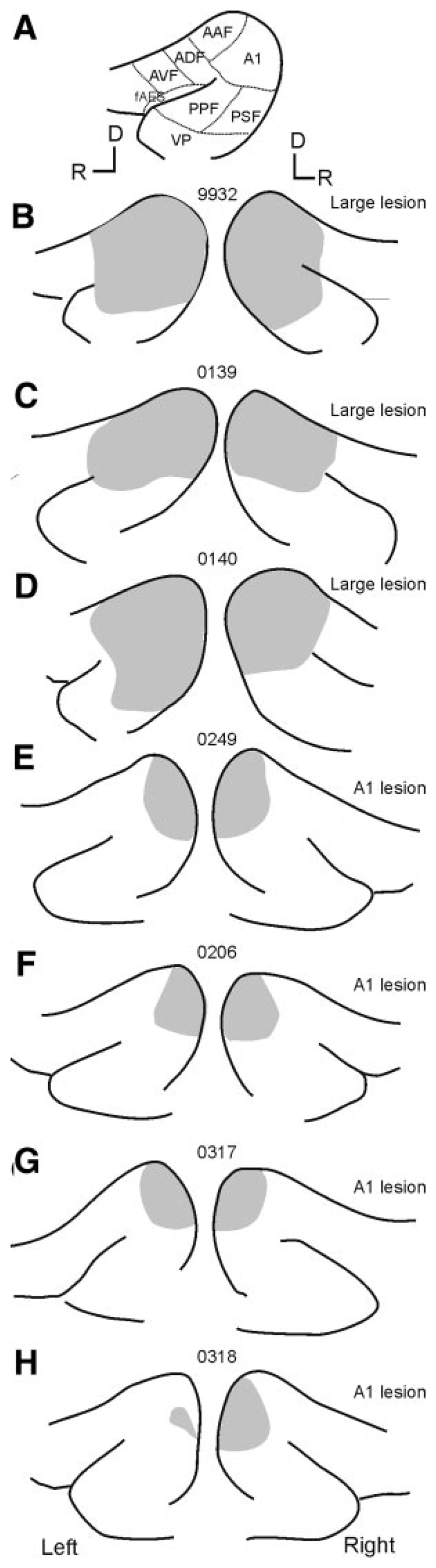
Schematics showing the size and location of the aspiration lesions made in each animal. In each case the suprasylvian sulcus and the pseudosyl-vian sulcus are shown with the lesion shaded in gray. A: schematic showing the location of auditory cortical fields (abbreviations as in Fig. 4). B–D: animals F9932, F0139, and F0140 received “large” lesions that included both the primary auditory fields and varying extents of the more ventral nonprimary fields. More restricted lesions were made in the other animals (E–H) after electrophysiological identification of the ventral low-frequency border of A1. D, dorsal; R, rostral.