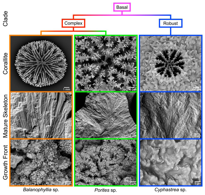
Figure 1.
Scanning electron microscopy (SEM) images of corallites from three distantly related coral genera: Balanophyllia, Porites, and Cyphastrea (top row). The bulk mature skeleton is always spherulitic (middle row), but the fine-scale morphology at the growth front varies widely across corals (bottom row).

