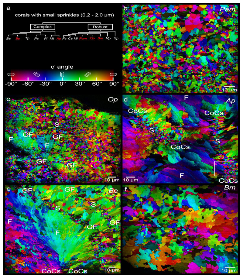
Figure 3.
PIC maps of the 5 coral species that exhibit abundant small (0.2-2μm) sprinkles across their skeletons. These are Phyllangia americana mouchezii (Pam), Oculina patagonica (Op), Acropora pharaonis (Ap), Balanophyllia europaea (Be), and Blastomussa merleti (Bm). Sprinkles (S) appear as randomly colored and oriented crystals smaller than fiber (F) crystals. In Ap they are localized in centers of calcification (CoCs, between pairs of CoC labels), in Op and Be at the surfaces of fiber bundles, which during coral skeleton growth were the growth fronts (GF, between pairs of GF labels). In Pam and Bm sprinkles appear everywhere interspersed with fibers. The box in d is magnified in Figure 6d.

