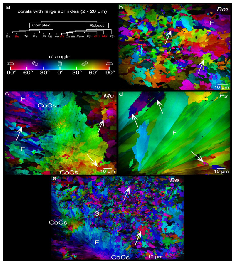
Figure 4.
PIC maps of the 4 coral species that exhibit large (2-20μm) sprinkles across their skeletons. These are Blastomussa merleti (Bm), Madracis pharensis (Mp), Favia sp. (Fs), and - Balanophyllia europaea (Be). Arrows indicate a few large sprinkles, but many more are visible. Large sprinkles are distinct from fibers (F) not by size but by crystal orientations: they form >35° angles with their neighboring crystals, whereas fiber crystals only form small angles (<35°) with respect adjacent fibers in the same bundle. Nanoparticulate crystals (between pairs of CoC labels) are visible in the CoCs of Mp and Be. Nanocrystals in the CoCs are not randomly oriented, but rather oriented similarly to their neighboring crystals. CoCs, therefore, cannot be sprinkles.

