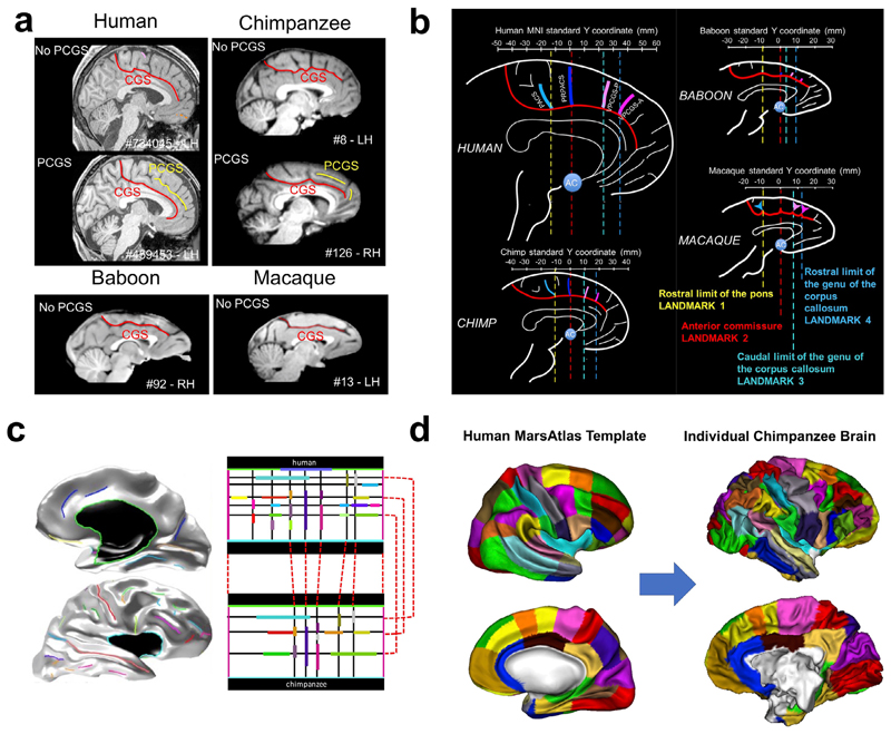
Fig. 3.
Sulcal anatomy for inter-primate brain comparisons. a) Emergence of the para-cingulate sulcus (PCGS) the primate medial frontal cortex (Amiez et al., 2019 : non-existing in baboons and macaques, but sometimes present for great apes and humans. b) Sulcal landmarks in the primate medial frontal cortex (Amiez et al., 2019). c) Projection of human brain sulci (left) onto a rectangular sulcal model (top right). Correspondences are defined between the human rectangular cortical sulci model and its chimpanzee equivalent (bottom right). d) Application of the model correspondences to map a human surface-based brain atlas onto an individual chimpanzee surface (Coulon et al., 2018).

