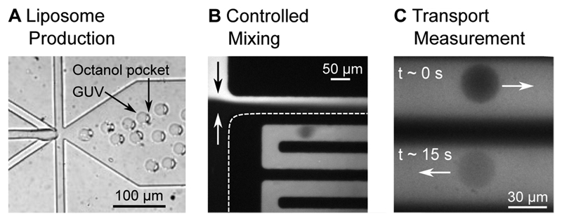
Figure 2.
Liposomes at different positions in the microfluidic chip. A: Liposome assembly at the 6 way formation junction. A 1-octanol pocket is initially attached to the liposomes. The liposome and the octanol pocket separate further downstream in the post-junction channel. B: The liposomes experience a spontaneous exposure to a drug solute at a T-junction where the two flows mix in a controlled manner. C: The liposomes, surrounded by the autofluorescing drug (λex = 350 nm), can be monitored at different parts of the channel, corresponding to different times that the liposome has been exposed to the drug. The increase in liposome intensity as the fluorescing drug diffuses across the membrane is used to determine the permeability coefficient of the drug across the lipid membrane under investigation.

