Fig. 4.
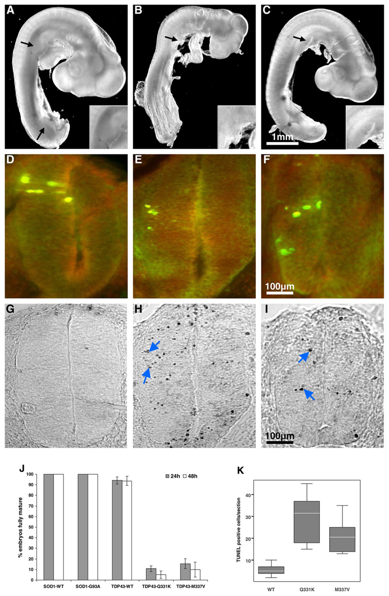
Mutant TDP-43 causes chick embryonic developmental delay. Chick embryo spinal cords were transfected with plasmids encoding wild-type TDP-43 (A, D, G), mutant Q331K (B, E, H) or M337V (C, F, I). Embryo images and sections shown were taken 24 hours post electroporation. Embryos expressing TDP43WT developed normal limb (black arrows) and tail buds (A) while those expressing mutant TDP-43 did not (B, C). Magnified images of upper limb buds are shown in insets. (J) The percentage of embryos that matured normally (reached HH stage 15 or 17 after 24 hours and 48 hours respectively) is shown. Data were generated from analysis of 49 embryos in each treatment group. (D-F) Transverse sections of chick spinal cord double-stained with anti-HA (green) and anti-myc (red) antibodies to the tags on transfected TDP-43 constructs. Unilateral TDP-43 construct expression in spinal cord neural cells occurs following embryo electroporation. The majority of the cells in the transfected region have the characteristic phenotype of neuroepithelial cells. (G-I) TUNEL staining of sections shown in D-F demonstrated apoptotic nuclei (blue arrows). (K) Quantification of apoptosis. TUNEL positive cells in five sections per embryo (two embryos per transfection) were counted.

