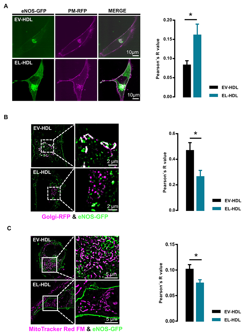
Fig. 2.
Impact of EL-HDL on subcellular localization of eNOS-GFP. EA.hy 926 cells overexpressing eNOS-GFP and (A) plasma membrane-RFP (PM-RFP) or (B) Golgi-RFP, or (C) stained with MitoTracker® Red were incubated with 100 μg/mL EV-HDL or EL-HDL for 16 h followed by (A) confocal microscopy or (B, C) structured illumination microscopy. Representative images and the Pearson's co-localization coefficients are shown. The colocalizations of eNOS-GFP (green) with (A) PM-RFP (magenta), (B) Golgi-RFP (magenta) or (C) MitoTracker® Red FM (magenta) are indicated in white. The values are expressed as mean ± SEM of 4 independent experiments performed in duplicate and analyzed by two-tailed unpaired t-test; *p < 0.05.

