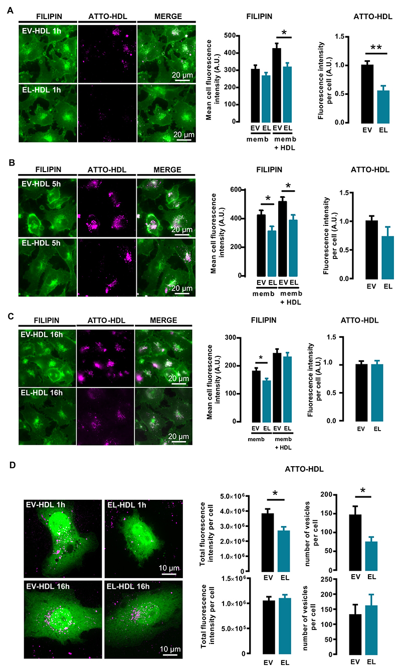
Fig. 3.
ATTO-EL-HDL uptake and impact on cellular FC content. EA.hy 926 cells were incubated with 100 μg/mL ATTO-EV-HDL protein or ATTO-EL-HDL protein for (A) 1, (B) 5 or (C) 16 h followed by washing, fixation, filipin staining, and imaging of filipin (green)- and ATTO-fluorescence (magenta) by wide-field microscopy. Representative images and corresponding analyses are shown. Total filipin-fluorescence (memb. + HDL) is the sum of the filipin fluorescence associated with cell membranes (memb.) and the filipin fluorescence of ATTO-HDL-vesicles (HDL), the latter determined by using the ATTO-fluorescence as a mask for the filipin fluorescence. The values are presented as mean ± SEM of 3 independent experiments analyzed by two-tailed unpaired t-test; *p < 0.05, **p < 0.01. (D) EA.hy 926 cells overexpressing the cytosolic marker geNOP-GFP were incubated with 100 μg/mL ATTO-EV-HDL protein or ATTO-EL-HDL protein for 1 or 16 h, followed by 3D spinning disk confocal microscopy to analyze the total ATTO-fluorescence and the number of ATTO-HDL-containing vesicles taken up per cell. Representative images and corresponding analyses are shown. The values are expressed as mean ± SEM of 4 independent experiments performed in duplicate and analyzed by twotailed unpaired t-test; *p < 0.05.

