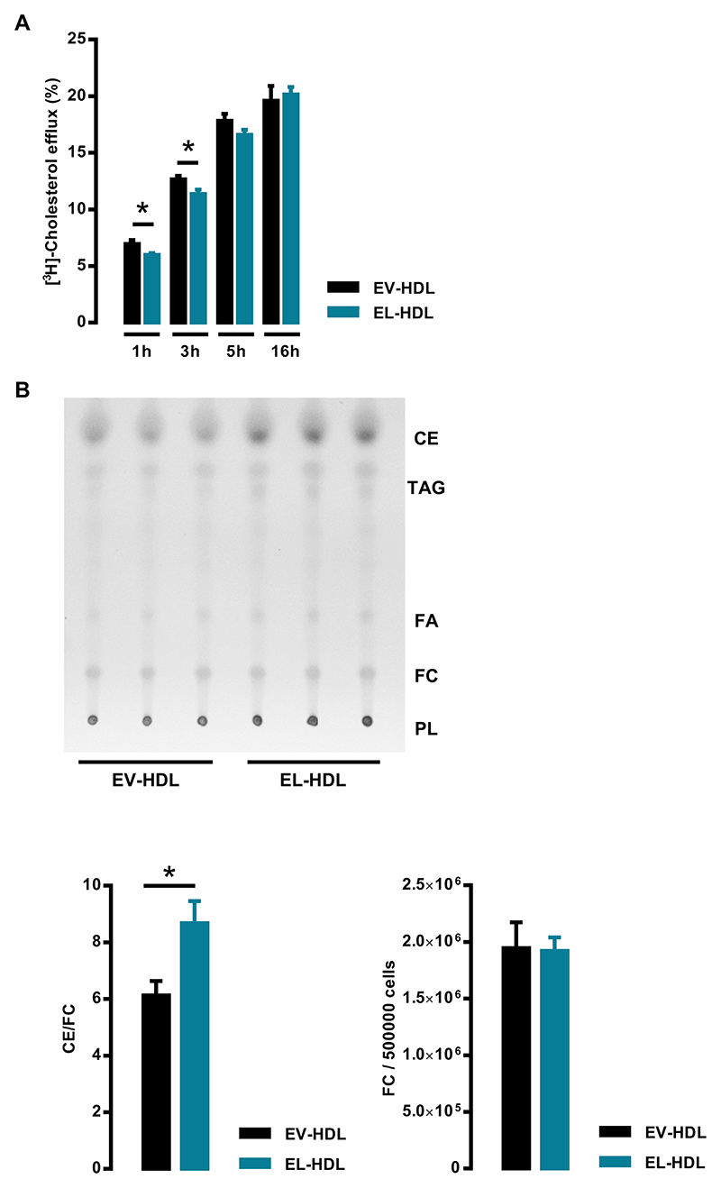
Fig. 4.
Impact of EL-HDL on cholesterol efflux as well as FC and CE content in EA.hy 926 cells. (A) Cells labeled with [3H]-cholesterol for 24 h were incubated with 100 μg/mL EV-HDL protein or EL-HDL protein for indicated time periods. Cholesterol efflux was expressed as the radioactivity in the medium relative to total radioactivity in medium and cells. The values are presented as mean ± SEM of 4 independent experiments performed in duplicate analyzed by two-tailed unpaired t-test; *p < 0.05. (B) EA.hy 926 cells plated in 6-well plates were incubated with 100 μg/mL of EV-HDL protein or EL-HDL protein for 16 h. Thereafter, the cells were washed with PBS and lipids were extracted with hexane/isopropanol (3:2, v:v). Extracts were evaporated and dissolved in chloroform before thin layer chromatography using hexane-diethyl ether-glacial acetic acid (70:29:1, v:v:v) as a mobile phase. The signals corresponding to phospholipids (PL), free cholesterol (FC), triacylglycerols (TAG), fatty acids (FA), and cholesterol ester (CE) were visualized by primulin and the signal intensity determined by densitometry. Annotations of the lipid species refer to the lipid standards on the plate. Representative TLC plate and the ratio of CE and FC signals as well as arbitrary units of FC per 500,000 cells both normalized to background are shown. The values are presented as mean ± SEM of 4 independent experiments performed in triplicate and analyzed by two-tailed unpaired t-test; *p < 0.05.

