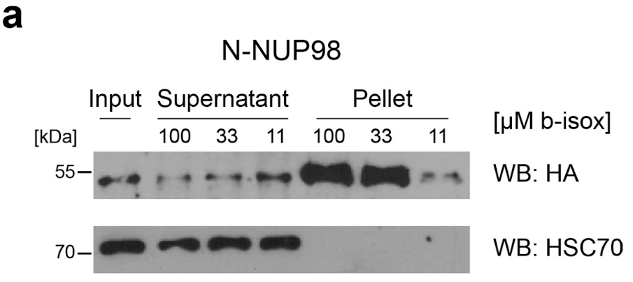
Extended Data Fig. 4. b-isox precipitation of N-NUP98, related to Fig. 4.

a, Western blot analysis of protein lysates from HEK293T cells expressing
N-NUP98 treated with 11 μM, 33 μM or 100 μM b-isox. N-NUP98 was detected using anti-HA antibodies. Total input, supernatant and b-isox fractions (pellet) are shown. Blot is representative of three independent experiments. Uncropped images are available in Supplementary Fig. 1.
