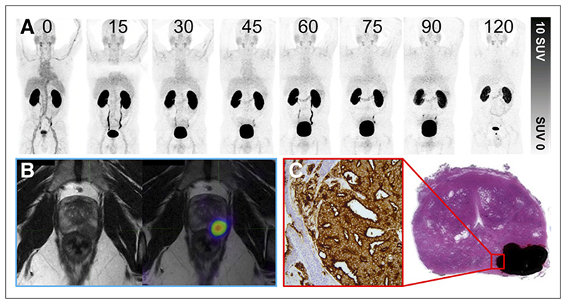
Figure 3. Patient with prostate adenocarcinoma with Gleason score of 4 + 4 = 8 and PIRADS 5 on prior multiparametric MRI.
(A) 68Ga-THP-PSMA PET multi-time-point images (0–120 min) demonstrating rapid blood-pool clearance and low background activity. (B) PET image (120 min) after voiding fused to MRI T2-weighted sequence. Focal uptake can be seen in left posterior midzone lesion. (C) Histopathologic correlation after prostatectomy. Area of adenocarcinoma is shaded in black; magnified area demonstrates 3+ staining on PSMA immunohistochemistry.

