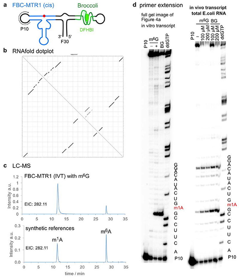
Figure 7. Extended Data Figure 7.
Plasmid-encoded cis-active ribozyme. a. Schematic depiction of F30-Broccoli-cis (FBC)-MTR1 construct. b. Dot plot for FBC-MTR1 generated by Vienna RNAfold (http://rna.tbi.univie.ac.at/), indicates high probability of folding into the designed structure. c. LC-MS analysis of SVPD/BAP-digested FBC-MTR1 in vitro transcript after reaction with m6G. Extracted ion chromatogram (EIC) for detection of MH+ 282.11±0.05 (methylated adenosines) shows production of m1A, and m6A to a small extent (due to partial Dimroth rearrangement during digestion). Bottom trace for synthetic references m1A and m6A (50 nM each), is same as shown in Fig 3d. d. Primer extension stop assays also confirm activity of FBC-MTR1 transcribed in vitro and in vivo, in the presence of total E.coli RNA. Left: full gel image shown in Fig. 4a for in vitro transcribed FBC-MTR1. Right: primer extension on total E.coli RNA, isolated 1 h after IPTG induction, and incubated with indicated m6G or BG concentration in vitro. These experiments were independently repeated two times with similar results.

