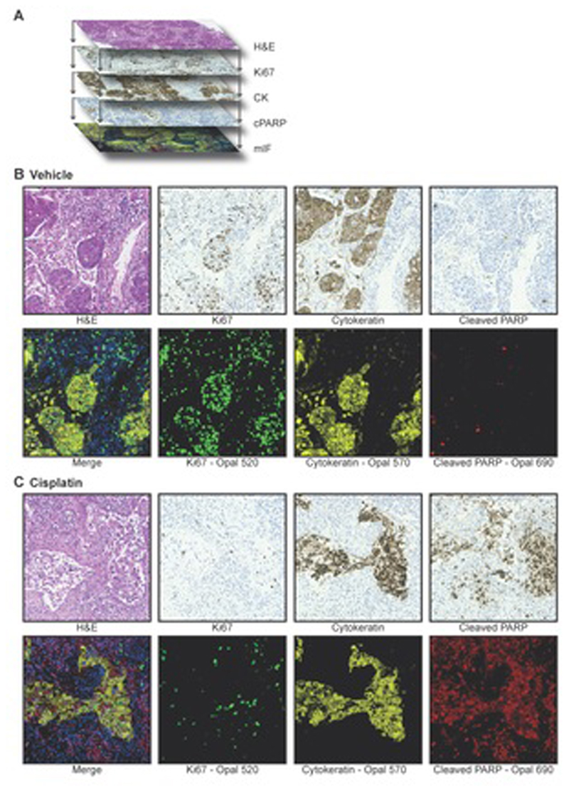
Figure 1. Immunostaining of NSCLC PDEs.
a Serial sections and sequential staining of parallel sections with: H&E, Ki67 using IHC, CK using IHC, cPARP using IHC and all three biomarkers together with DAPI by mIF.
b and c Serial sections from vehicle (b) or cisplatin (c) treated NSCLC PDEs of the lung adenocarcinoma subtype were stained with H&E, with the three biomarkers (Ki67, CK, cPARP) on sequential sections using IHC, or with the three biomarkers on the same section using mIF. The merged image generated from mIF shows integration of Ki67 (green), CK (yellow), cPARP (red) with DAPI (blue) staining.

