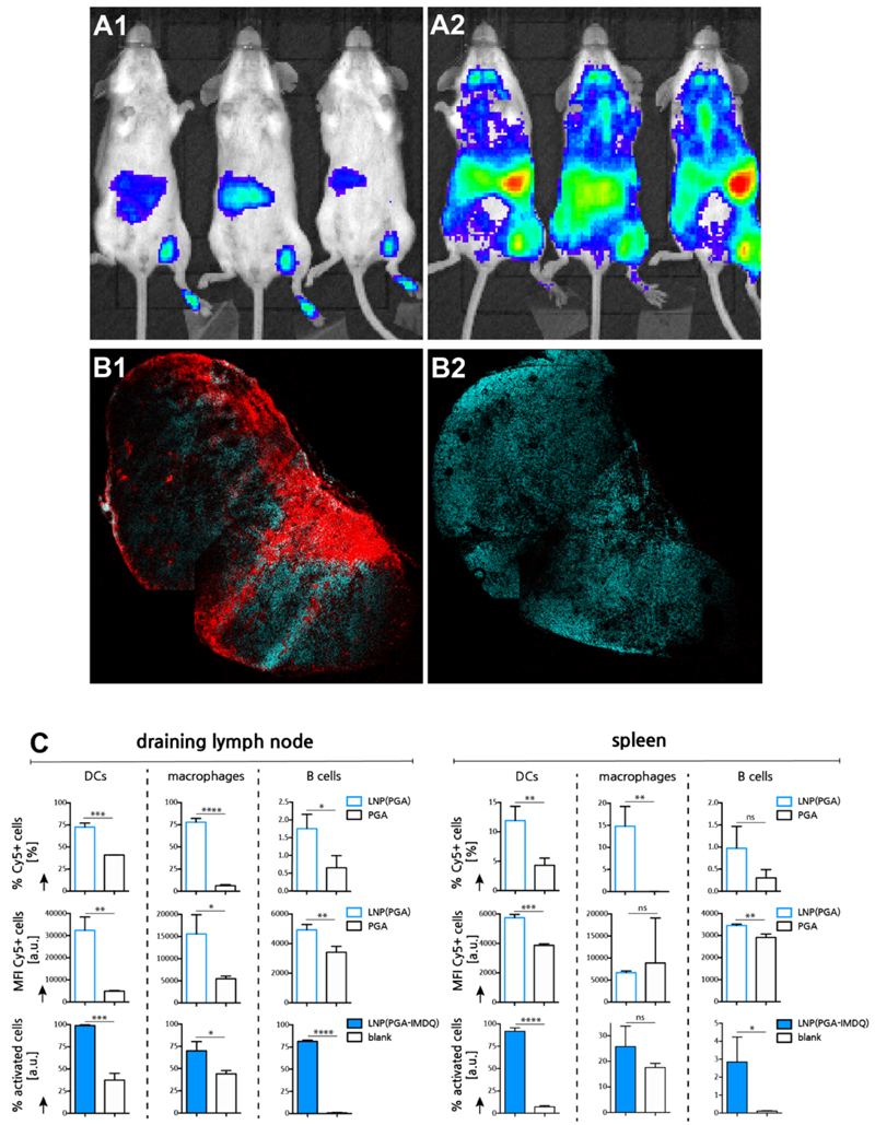
Figure 5. In vivo lymphatic transportation and innate immune activation by LNP.
(A) Bioluminescence imaging of IFNb-luciferase reporter mouse 4h post injection. (A1: PGA-IMDQ; A2: LNP(PGA-IMDQ)) (B) Confoca microscopy image of a sectioned popliteal lymph node, 24h post injection of LNP in the foot pad. (cyan: DAPI, red:Cy5-PGA loaded LNP). Note that some irregularities in the image are due to stitching. (B1 : LNP(Cy5-PGA); B2: Cy5-PGA) (C) Flow cytometry analysis of LNP uptake by innate immune cells and innate immune cell activation in the draining inguinal lymph node) 24h post injection of LNP in the tail base (n=3; t-test *: p<0.05, **:p<0.01, ***:p<0.001, ****:p<0.0001).

