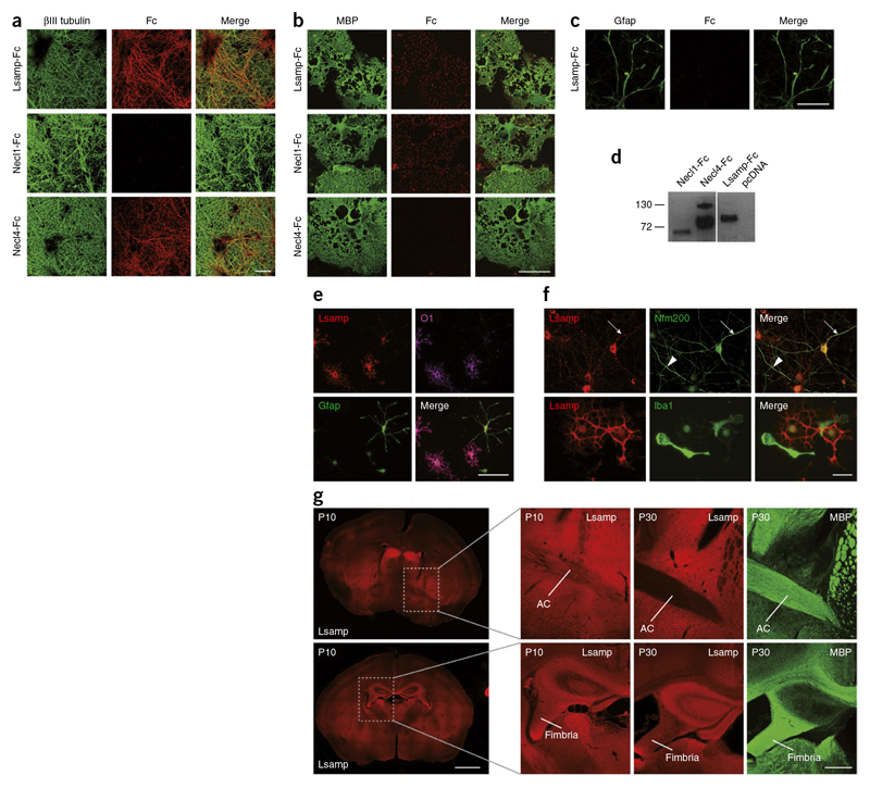
Figure 7. Lsamp interacts with and is expressed on oligodendrocytes and neurons.
(a) Binding of Lsamp-Fc to neurons labeled with an antibody against βMI tubulin. Necl4-Fc served as a positive and Necl 1-Fc as a negative control.The Fc fragments were visualized with Cy3-conjugated anti-Fc antibodies. (b) Binding of Lsamp-Fc to oligodendrocytes labeled with an antibody against MBP. Necl 1-Fc served as a positive and Necl4-Fc as a negative control. (c) Lsamp-Fc did not bind to astrocytes labeled with an antibody against Gfap. (d) Immunoblot of secreted Fc fusion proteins containing the extracellular domains of the indicated proteins. (e) Immunofluorescence of mixed glial cultures shows staining of oligodendrocytes (O1; magenta), but not astrocytes (Gfap; green) with an antibody against Lsamp (red). (f) Top, Lsamp was present on a subpopulation of neurons (neurofilament 200 kDa) (top right). Arrow indicates toward an Lsamp-positive neuronal process, arrowhead indicates an Lsamp-negative process. Bottom, Lsamp was absent from microglia (Iba1), as shown by staining of a mixed glial culture. (g) Brain sections of wild-type mice were immunostained with antibodies against Lsamp (red) at P10, and Lsamp and MBP (green) at P30. Lsamp staining was enriched along the axonal tracts of the fimbria and anterior commissure (AC) at P10, but not at P30. Scale bars represent 10 μm.

