
Figure 6.
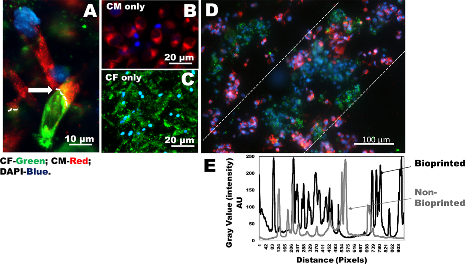
Heterocellular coupling of CM and CF. (A) Human CM (prestained using PKH26, red) and CF (prestained using PKH67, green) coupled in 2D wells in the ratio of CM/CF (1:1) shown by white arrows and dotted lines. (B, C) Images of controls for either CM or CF (in 2D wells). (D) Results of heterocellular coupling between prestained CM (red) and CF (green) from 3D bioprinted constructs. Nonprinted controls that included cells mixed in gels are shown in Figure S5. The white dashed lines in (D) denote that the cells are confined to their printed patterns. However, these samples were fixed prior to DAPI staining, and the cells were not viable during imaging. To confirm heterocellular coupling using viable cells, we report results in Figure 7. (E) Intensity values within the white dotted lines in a bioprinted sample (black line) versus a nonbioprinted sample (gray line). The average intensity values for the bioprinted samples exhibited more frequent peaks with respect to distance, depicting a higher frequency of cells present in the preferred orientation compared to samples that were nonbioprinted.