
Fig. 5.
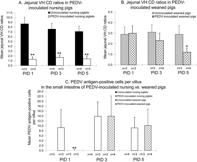
Mean ratios of jejunal villous height to crypt depth (VH:CD) in PEDV-inoculated nursing (A) and weaned (B) pigs. (C) Mean PEDV antigen-positive cells per villus in the small intestine of PEDV-inoculated nursing piglets vs. weaned pigs. Eight pieces of formalin-fixed mid- to distal jejunum were taken from each virus-infected and control pigs for morphometric analysis. Only well-orientated, H&E- or IHC-stained jejunal sections were measured. Villous height, crypt depth, and number of PEDV antigen-positive cells were estimated by measuring at least 10 villi and crypts throughout the section. Each bar represents the mean ± SEM. *, P < 0.05; **, P < 0.01 (statistically significant differences between the PEDV-inoculated nursing and weaned pigs by Student’s t-test).