
Fig. 2.
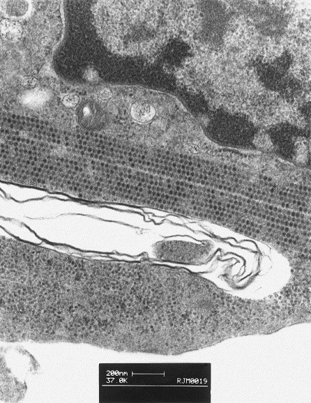
Transmission electron micrograph of Crandell feline kidney cells infected with FCV-Ari. Parallel stacked linear arrays made up of numerous calicivirus virions 25–30 nm in diameter are present in the cytoplasm of infected cells.
Official websites use .gov
A
.gov website belongs to an official
government organization in the United States.
Secure .gov websites use HTTPS
A lock (
) or https:// means you've safely
connected to the .gov website. Share sensitive
information only on official, secure websites.
Transmission electron micrograph of Crandell feline kidney cells infected with FCV-Ari. Parallel stacked linear arrays made up of numerous calicivirus virions 25–30 nm in diameter are present in the cytoplasm of infected cells.