
Fig. 5.
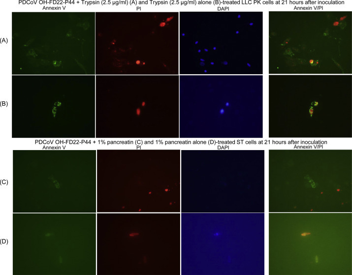
Annexin V staining in the LLC porcine kidney (LLC-PK) cells (A and B) and swine testicular (ST) cells (C and D) inoculated with the cell-adapted PDCoV strain OH-FD22 (virus passage number, 44), as supplemented with 2.5 μg/ml of trypsin or 1% pancreatin in cell culture medium. (A) Green-fluorescent annexin V, red-fluorescent propidium iodide (PI), and blue-fluorescent 4′, 6-diamidino-2-phenylindole dihydrochloride (DAPI) staining of the inoculated LLC-PK cells at 21 h after inoculation, showing a small number of annexin V+/PI+/DAPI+ cells. (B) Green-fluorescent annexin V, red-fluorescent PI, and blue-fluorescent DAPI staining of non-inoculated, trypsin (2.5 μg/ml) only-treated LLC-PK cells at 21 h after inoculation, showing few annexin V+/PI+/DAPI+ cells. (C) Green-fluorescent annexin V, red-fluorescent PI, and blue-fluorescent DAPI staining of the inoculated ST cells at 21 h after inoculation, showing a small number of annexin V+/PI− or annexin V−/PI+ cells. (D) Green-fluorescent annexin V, red-fluorescent PI, and blue-fluorescent DAPI staining of non-inoculated, 1% pancreatin only-treated ST cells at 21 h after inoculation, showing few annexin V+/PI+ cells. Original magnification, all ×600. (For interpretation of the references to color in this figure legend, the reader is referred to the web version of this article.)