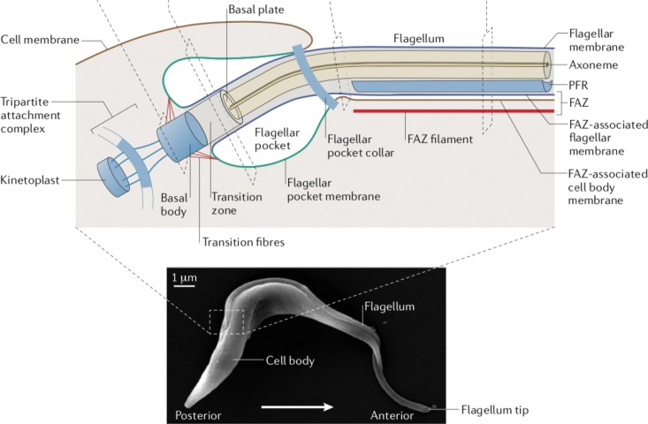
FIG 1.
Diagram of the flagellum and surrounding structures in BF T. brucei. The image on the bottom is a scanning electron micrograph of a BF trypanosome. The section in the dashed box, representing the base of the flagellum and the surrounding cellular components, has been expanded as a schematic diagram at the top of the figure. Subcellular structures associated with the flagellum are indicated by labeling. PFR indicates the paraflagellar rod and FAZ the flagellum attachment zone. (Adapted from reference 6 with permission of Springer.)

