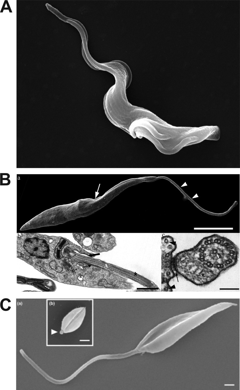
FIG 2.
Images of kinetoplastid parasites and their flagella. (A) Scanning electron micrograph of bloodstream form Trypanosoma brucei. The parasite has begun cell division, and the flagellum has duplicated at the posterior end of the cell (lower right). The anterior tip of the flagellum that is unattached to the cell body can be seen in the upper left. (Reproduced from the Wellcome Collection [https://wellcomecollection.org/works/t3za8n35]; Gull Laboratory, courtesy of Sue Vaughan.) (B) Electron micrographs of epimastigote forms of Trypanosoma cruzi. (a [top]) Scanning electron microscopy image with an arrow pointing to the flagellar pocket and two arrowheads pointing to the region of the flagellum that is free from the cell body; (b [lower left]) TEM image with a thick arrow pointing to the flagellar pocket and a thin arrow pointing to the flagellum attachment zone (FAZ); (c [lower right]) TEM image transverse to the flagellum, with the arrow pointing to the FAZ (connecting the flagellar and cell body membranes) and the flagellar membrane visible as a dark contour surrounding that organelle. (Reproduced from reference 131.) (C) Scanning electron microscopy images of an L. mexicana promastigote (a) and amastigote (b, inset). The arrowhead in subpanel b points to the short amastigote flagellum. (Reproduced from reference 132 with permission of Elsevier.)

