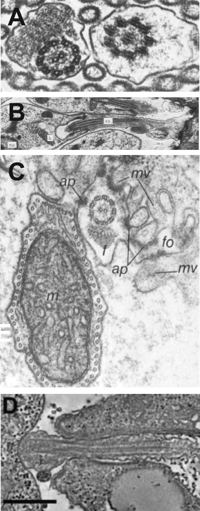
FIG 3.
Images of flagella from kinetoplastid parasites interacting with tissues of insect vectors (A to C) or with internal organelles of host cells (D). (A) Transverse view of two flagella from nectomonads of L. amazonensis interacting with the midgut microvilli of a sand fly. (Reproduced from reference 23 with permission of the Royal Society of London.) (B) Flagellum of a haptomonad of L. amazonensis forming a foot-like adhesion (extreme right of image) with the stomodeal valve of a sand fly. Labels represent the flagellar axoneme (ax), the kinetoplast (k), and the nucleus (nu). (Reproduced from reference 23 with permission of the Royal Society of London.) (C) Flagellum (f) of a T. brucei epimastigote attached to salivary gland microvilli (mv) of a tsetse fly. Labels represent flagellar outgrowth (fo), attachment plaque (ap), and trypanosome mitochondrion (m). (Reproduced from reference 26 with permission of the Company of Biologists.) (D) Attachment of the tip of an amastigote flagellum to the parasitophorous vacuole membrane of a host macrophage. (Reproduced from reference 29 with permission of the Federation of American Societies for Experimental Biology.)

