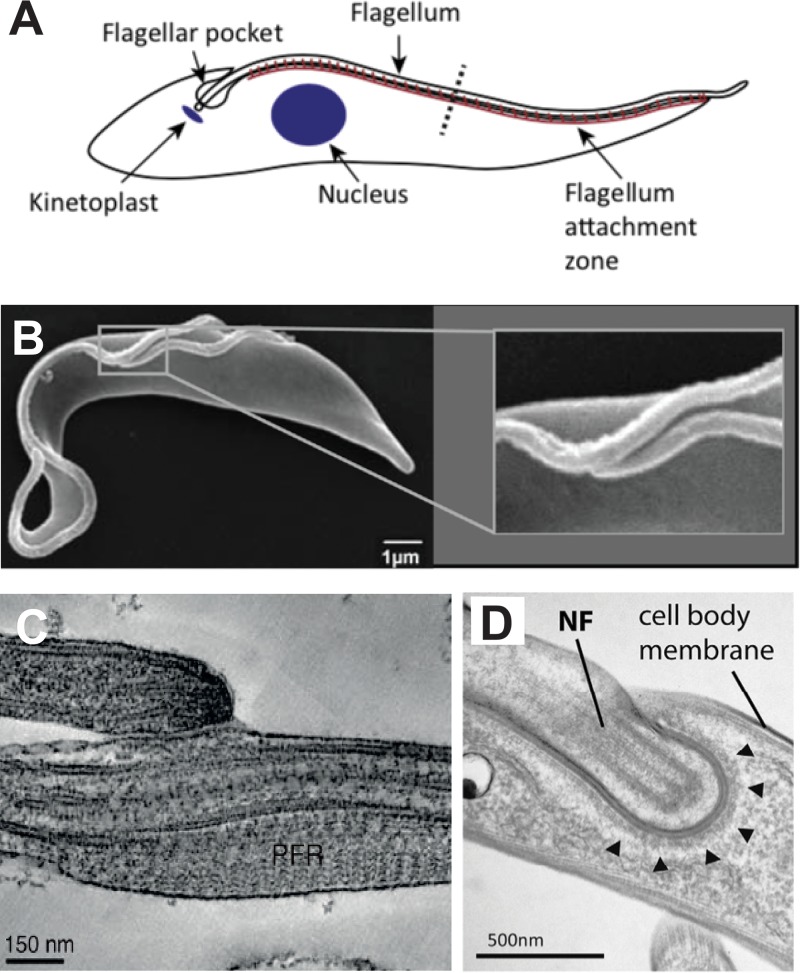
FIG 4.
Interactions of the T. brucei flagellum with other components of the parasite. (A) Schematic diagram of a BF parasite showing the flagellum attachment zone (brown line) attaching the flagellum to the cell body. (Reproduced from reference 32 with permission of Elsevier.) (B) Scanning electron micrograph of a replicating PF trypanosome showing the shorter new flagellum attached at its growing end to the longer old flagellum. The inset at the right shows an expanded view of the boxed region containing the two attached flagella. (Reproduced from reference 52.) (C) Transmission electron micrograph of the flagellum connector attaching the growing tip of the new flagellum (top) to the lateral membrane of the old flagellum (bottom). (Reproduced from reference 52.) (D) Transmission electron micrograph of the flagellar groove of a replicating BF trypanosome. NF indicates the tip of the new flagellum embedded in a groove within the cell body membrane. Arrowheads indicate electron dense material lining the cytosolic side of the groove membrane. (Reproduced from reference 55 with permission of the Company of Biologists.)

