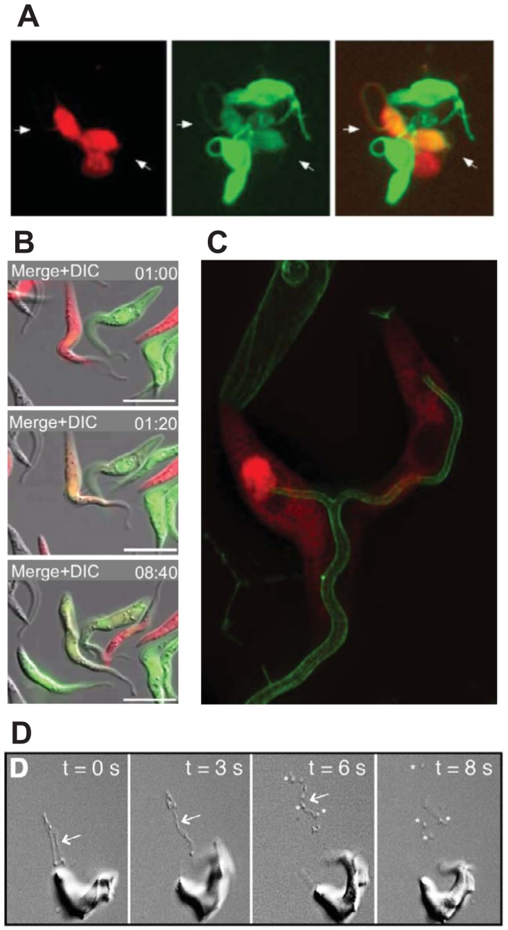
FIG 5.

Flagellar interactions in T. brucei. (A) Gametes of insect-derived trypanosomes labeled with either red or green fluorescent proteins interact via their flagella (two parasites indicated by white arrows) and exchange their cytosolic contents, resulting in dually red and green labeled parasites. (Reproduced from reference 59.) (B) Time-lapse fluorescence microscopy of mixed red and green fluorescently labeled PF trypanosomes (top panel, 1:00 min) shows that some parasites interact via their flagella and exchange cytosolic contents, producing dually labeled (orange) parasites (middle panel, 1:20 min). (Reproduced from reference 63.) (C) Structured illumination fluorescence microscopy shows two PF parasites labeled in the cytosol with DsRed (red) and on the flagellar membrane with calflagin44-GFP (green). The two flagella have fused to form a single wider flagellar structure (bottom of the image). (Reproduced from reference 63.) (D) Video microscopy of differential interference images of BF T. brucei show the emergence of nanotubes (arrows) from the flagellar membrane (0 to 6 s), followed by fragmentation into extracellular vesicles (asterisks, 6 and 8 s). (Reproduced from reference 73 with permission from Elsevier.)
