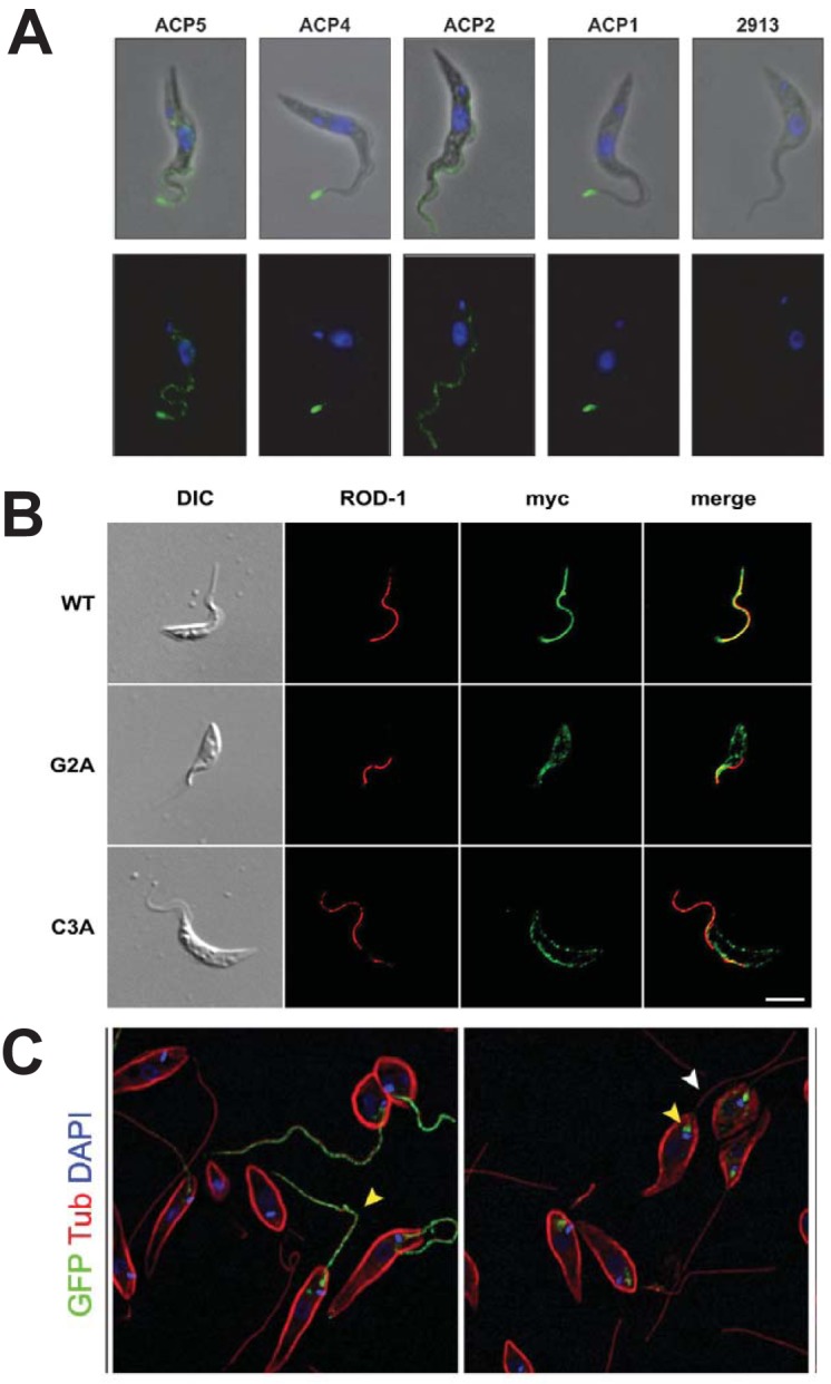
FIG 6.

Flagellar localization of several membrane proteins. (A) Flagellar localization of 4 hemagglutinin (HA)-tagged adenylate cyclase proteins (ACPs) in PF T. brucei. Green fluorescence indicates localization of the relevant HA-fusion proteins, and blue is DAPI (4′,6′-diamidino-2-phenylindole) fluorescence from nuclear and kinetoplast DNA. The top panels are superpositions of phase-contrast and immunofluorescence images, and the bottom panels are immunofluorescence images. The panels marked 2913 represent a parasite not expressing any HA fusion protein. (Reproduced from reference 78 with permission.) (B) The location of calflagin Tb44 in PF T. brucei was monitored using antibody against the myc epitope tag (green) and antibody against an endogenous paraflagellar rod protein (ROD-1, red). Wild-type (WT) Tb44 colocalized with ROD-1 to the flagellum, whereas the Tb44 mutants G2A (nonacylated) and C3A (nonpalmitoylated) did not localize to the flagellum. (Reproduced from reference 133 with permission from the Company of Biologists.) (C) Flagellar localization of GT1-GFP (green, yellow arrowhead) was observed in wild-type L. mexicana promastigotes (left panel), but the fusion protein arrested in the flagellar pocket (yellow arrowhead, right panel) in the Δkharon mutant, showing dependency of flagellar trafficking upon the LmxKHARON protein. Red fluorescence represents staining with anti-α-tubulin, which stains the subpellicular microtubules and flagellar axoneme (white arrowhead). (Reproduced from reference 111 with permission of the American Society for Biochemistry and Molecular Biology.)
