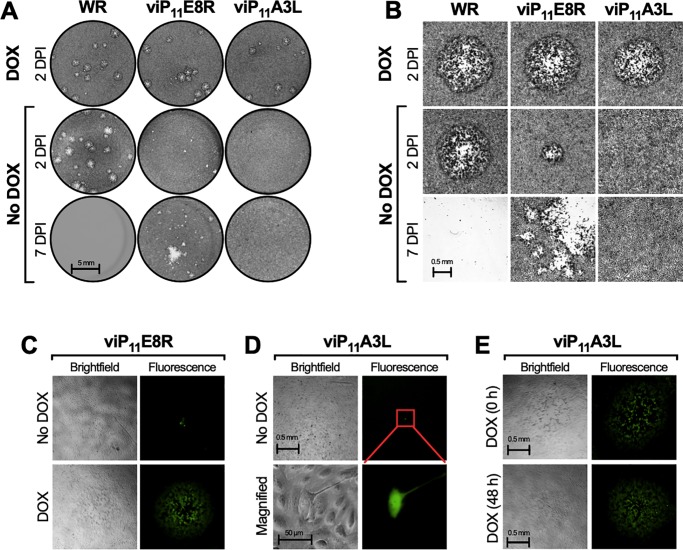
Fig 4. viP11A3L does not form plaques and causes abortive infections in the absence of DOX.
BS-C-1 cell monolayers were infected with the indicated VACVs at approximately 5–20 PFU/well in the absence or presence of 1 μg/ml DOX and cells were stained with crystal violet 2 or 7 DPI (A and B) or imaged by brightfield (phase) and fluorescence microscopy (C, D, and E). (A) Image of representative wells showing the plaque phenotypes. (B) Representative brightfield microscopic images of stained cells showing plaques, when present. (C) In the absence of DOX smaller plaques formed 2 DPI with viP11E8R. (D) In the absence of DOX, EGFP expression was contained to single viP11A3L-infected cells and was the only indication of infection. (E) When DOX was added at the time of infection or 48 h after infection, plaques were visible 2 and 4 days later, respectively. Data is representative of two separate experiments.

