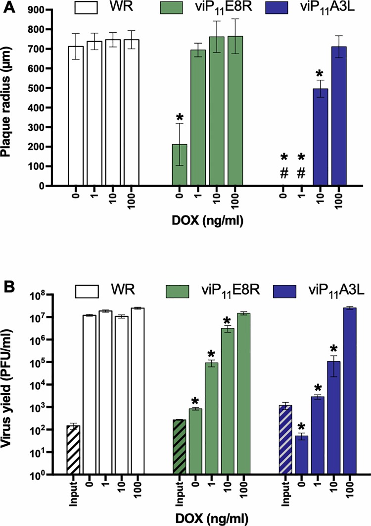
Fig 5. viP11A3L replicates indistinguishably from wild-type VACV in the presence of DOX.
(A) The effect of DOX on plaque size was examined by infecting BS-C-1 cell monolayers with the VACVs in the absence or presence of multiple concentrations of DOX. At 36 hpi, cells were stained with crystal violet and the size (radius) of approximately 20 representative isolated plaques was measured (# indicates absence of plaques). (B) The effect of DOX on virus replication was examined by infecting BS-C-1 cell monolayers with the indicated VACVs at an MOI of 0.01. Cells were collected immediately to determine input titer (hatched bars) or after 48 h in the absence or presence of multiple concentrations of DOX to determine virus yield (solid bars). Titers were determined on BS-C-1 cells in the presence of 1 μg/ml DOX. The data shown represent the mean viral yields from triplicate samples assayed in duplicate. Error bars indicate standard deviation. An asterisk indicates statistically significant differences (p < 0.05 by two-way ANOVA followed by Tukey’s multiple comparisons test) between WR and the inducible viruses at a given DOX concentration.

