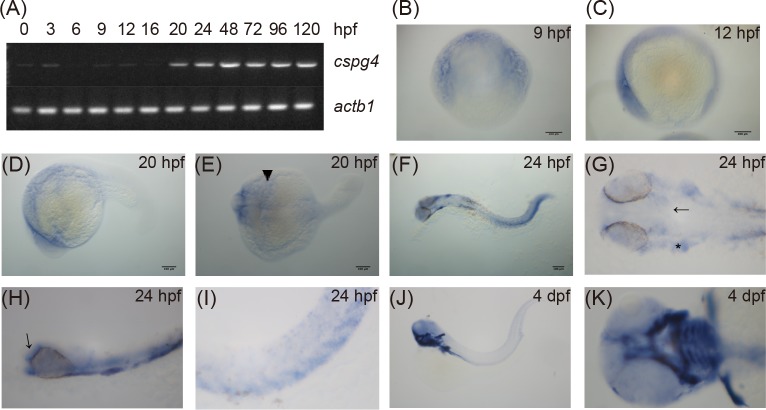
Fig 2. Zebrafish embryo expressed cspg4 during development.
(A) The expression of cspg4 was detected by RT-PCR from 0 to 120 hpf with actb1 as loading control. (B-H) Expression pattern of cspg4 was revealed by whole-mount in situ hybridization. At 9 (B) and 12 hpf (C), cspg4 was expressed in both the anterior and posterior end of the embryos. Lateral view (D) and dorsal view (E) of 20 hpf embryos showed that cspg4 was mainly expressed in the anterior paraxial and lateral mesoderm (arrow head). (F) Cspg4 was expressed in head, somite, ventral mesenchyme and tail at 24 hpf. Scale bar represents 100 μm. (G) Cspg4 was expressed in head mesenchyme (arrow) and otic placode (asteridsk). (H) Lateral view of 24 hpf embryo showed that cspg4 was expressed in the head mesenchyme (arrow) and pharyngeal region. (I) Cspg4 was expressed in the ventral sclerotome. (J) Cspg4 expressed in jaw, pectoral fins, and trunk at 4 dpf. (K) Ventral view of a 4 dpf larvae showed that cspg4 was expressed in pharyngeal cartilage and the pectoral fin.

