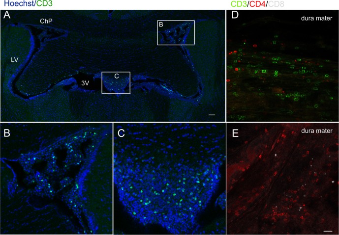
Fig 6.
The infiltration of T lymphocytes in the meninges and brain parenchyma after the adoptive transfer of BCG-induced T lymphocytes (A-E). Representative confocal micrographs of CD3+ lymphoid cells in the brain slice (A), the choroid plexus (ChP) (B) and the third ventricle (C). Numerous CD3+ and CD4+ lymphoid cells were found in the dura mater while few CD8+ lymphoid cells were found (D, E). Scale bar: 100 μm in A; 20 μm in B-E.

